


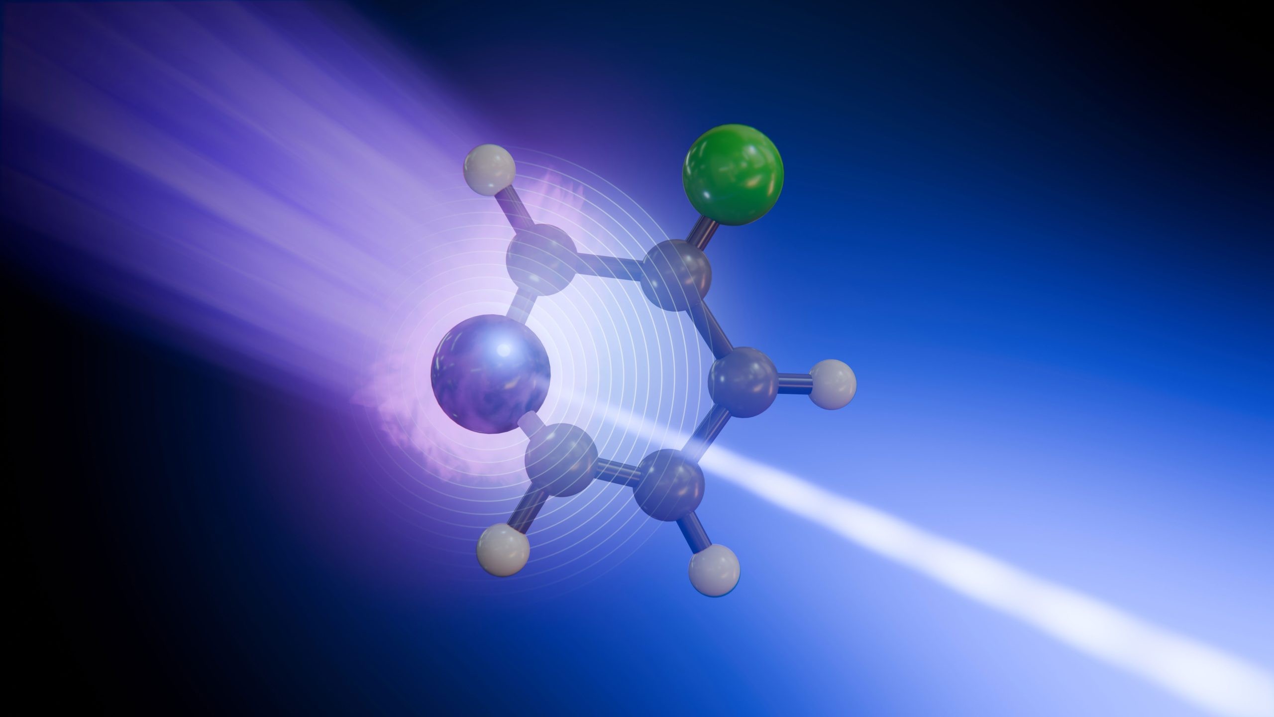
Chemistry is messy. It happens in trillionths of a second, a blur of shifting electrons and bending bonds that most instruments simply can’t catch.
But scientists at the European XFEL have managed to slow it down. By watching 3-fluoropyridine absorb light, they didn’t just see the reaction happen—they saw it unfold atom by atom. The catch? Not every atom tells the same story.
This isn’t just about speed. It’s about perspective.
Why nitrogen and fluorine react differently to light
Most people assume a molecule acts as a single, rigid unit when it reacts. Wrong.
In this study, researchers found that different atoms within the same molecule record different parts of a photochemical reaction. It turns out that tracking electronic and vibrational dynamics separately is only possible if you look in the right place.
They chose 3-fluoropyridile for a reason. It has a nitrogen atom and a fluorine atom. Two distinct observation points in one tiny ring.
Here is how the breakdown works:
- The fluorine atom acts as a clear indicator for vibrational relaxation. It shows how excess energy redistributes through the molecule’s motion.
- The nitrogen atom gets it more complicated. Because it participated directly in the initial excitation, its signal is a mix of changing electron distribution and structural movement
One watches the shake. The other watches the spark.
How time-resolved X-ray spectroscopy captures fleeting moments
You can’t photograph a chemical bond breaking with a camera. You need X-rays.
Specifically, you need time-resolved X-ray photoelectron spectroscopy (tr-XPS). The team used this method at the Small Quantum Systems (SQS) instrument at the European XFEL.
The process is a precise dance of light:
- An ultraviolet laser pulse hits the molecule. It absorbs the energy, electrons jump to a higher state, and the flat structure starts to distort.
- The molecule reaches a “conical intersection”—a fleeting junction where electronic states meet and energy transfers rapidly.
- A soft X-ray pulse follows after a controlled delay. It knocks out tightly bound electrons from either the nitrogen or fluorine atom.
The energy of those ejected electrons reveals the local chemical environment. By repeating this with varying delays, the team created a sequence of snapshots spanning just a few picoseconds.
“We can now see that not every atomic site tells us the same story in the signals we capture,” says co-author Antonio Picón. “Some atoms report where the charge goes, while others reveal how the molecule vibrates.”
Without advanced simulations, these energy shifts are just data. With them, they become a movie. The models connect the shifting electron energies to the physical movements of the nuclei, reconstructing how charge and motion develop over time.
What this means for studying DNA and solar materials
So why do we care about one specific ring of atoms?
Because this method separates electronic changes from structural motion even when they happen simultaneously. Previously, these two processes were often blurred together in measurements. Now, they can be pulled apart.
This capability opens doors for several fields:
- DNA protection: Understanding exactly how molecules dissipate energy to protect genetic code from light damage.
- Energy harvesting: Watching energy move through light-harvesting materials in solar tech.
- Biomolecular building blocks: Analyzing light-induced changes in complex biological systems.
Daniel Rivas, a co-author and former instrument scientist at SQS, puts it bluntly: “This is what European XFEL was built for—watching chemical change where it begins, on its natural timescale.”
It’s a new window into photochemistry. A microscopic one, but clear enough.
The challenge now isn’t just seeing the reaction. It’s knowing which atom to watch to get the truth.


